Gastrulation is one of the stages of early embryonic development through which the trilaminar germinative disc is formed, a structure with 3 differentiated embryonic layers that will give rise to all the organs and tissues of the embryo.
Gastrulation occurs just after segmentation and embryo implantation, i.e., when the embryo has already divided into many cells, has become a blastocyst and has managed to nest in the endometrium, initiating pregnancy.
Provided below is an index with the 8 points we are going to expand on in this article.
- 1.
- 2.
- 2.1.
- 2.2.
- 2.3.
- 3.
- 4.
- 4.1.
- 4.2.
- 4.3.
- 5.
- 6.
- 7.
- 8.
Beginning of embryonic development
After embryo implantation, between weeks 4 and 5 of pregnancy, the blastocyst-state embryo begins to proliferate rapidly and undergo changes in its structure.
The blastocyst is the state in which the embryo is from the fifth day of development, which has the structure of a blastula and it is possible to differentiate two cell types: the internal cell mass (ICM) and the trophectoderm.
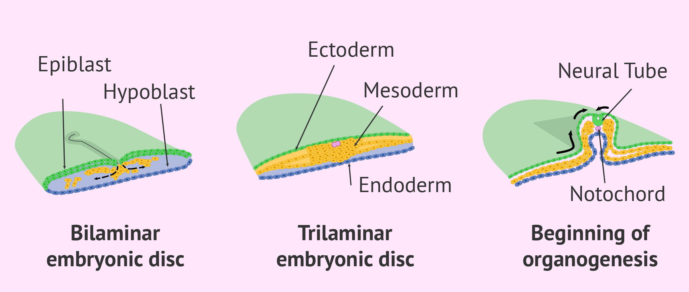
The internal cell mass of the blastocyst becomes a kind of flattened mass which is called embryonic discwhich is organized in two layers: the epiblast and the hypoblast.
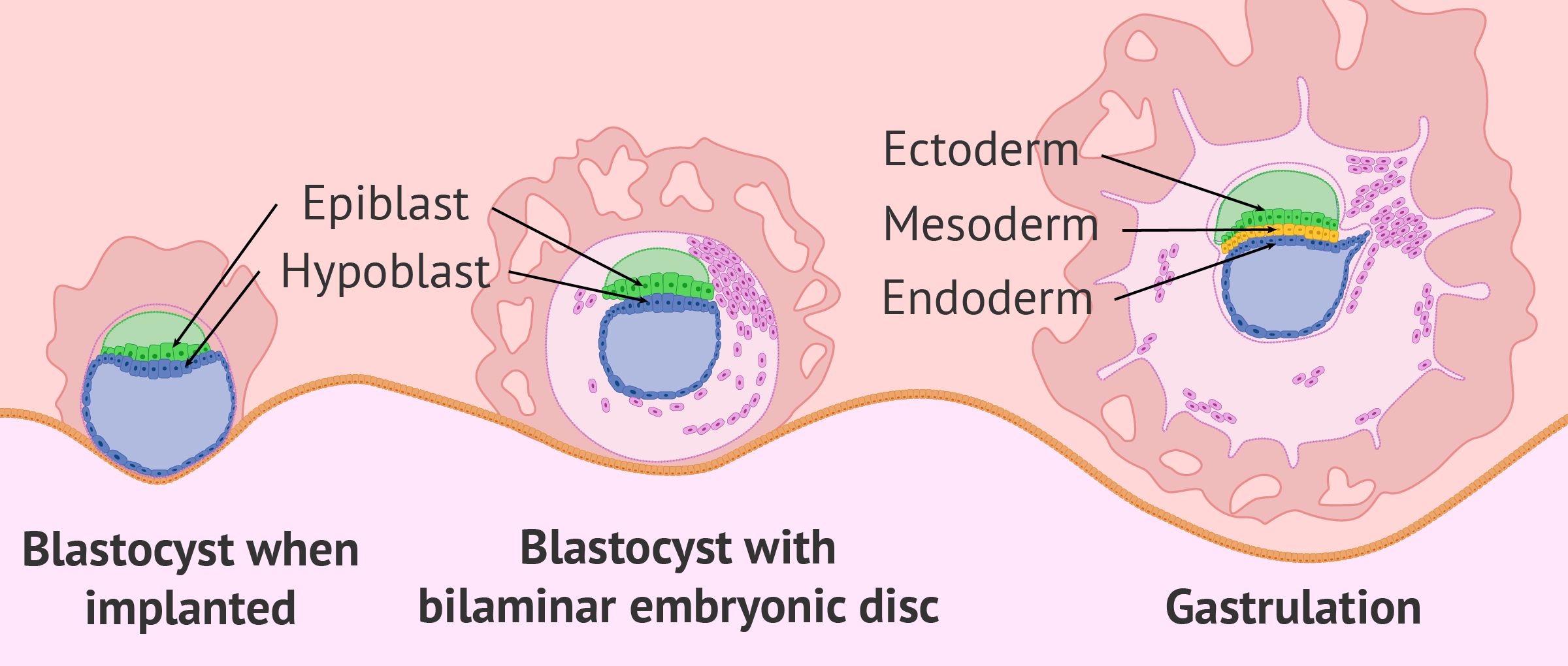
This embryonic disc is the origin of all the tissues and organs of the future embryo. On the other hand, the cells of the trophectoderm differentiate into other structures that will form the placenta.
How is gastrulation produced?
Gastrulation is the process by which the bilaminar embryonic disc becomes a trilaminar embryonic disc, with 3 different embryonic layers: ectoderm, mesoderm and endoderm.
Specifically, gastrulation starts from the blastula, when epiblastic cells divide and proliferate rapidly, so they need to migrate to new locations in the embryo.
This need for cellular migration causes the cells of the epiblasts to move towards the hypoblasts and displace the cells of the epiblasts, with which the third embryonic layer (mesoderm) appears between the two previous layers.
The process of human gastrulation is possibly the most important stage of embryonic development, since all tissues and organs of the body will be generated from the three embryonic layers or leaves.
Each one of them will be destined to form a different type of tissue, as we will comment next.
Ectoderm
It is the outermost layer surrounding the embryo and the first to form from the epiblasts.
With the beginning of organogenesis, the ectoderm cells will differentiate into two structures:
- The neural ectoderm
- will form the nervous system, that is, the brain and spinal cord.
- The surface ectoderm
- will form the most superficial tissues, such as the epidermis, hair, nails, mammary glands, subcutaneous glands and tooth enamel.
In addition, a group of ectodermal cells, the amniocytes, will form part of the amniotic sac where the embryo will be suspended when the amniotic fluid forms.
Mesoderm
It is the intermediate layer, but the one that is formed in the last place from the migration of the cells of the epiblasto, as we have commented.
The mesoderm is the embryonic leaf that will form most of the tissues and organs of the future fetus. In order to do this, we first differentiate in the following structures:
- The notochord
- is situated on the longitudinal axis of the embryo, from the base of the head to the tail, and acts as a support. The notochord is also fundamental for the formation of the neural tube from the ectoderm.
- Paraxial mesoderm
- develops on the back of the embryo along the notochord. Mesodermal cells form so-called pairs of somites, cell blocks on both sides of the neural tube that are responsible for forming muscle tissue, skeletal, cartilage and dermis.
- Intermediate mesoderm
- also known as nephrotome, as it will result in the kidneys on both sides of the embryo and other components of the urogenital system.
- Lateral mesoderm
- is the most external part of the mesoderm and the one that will originate the blood and cardiovascular system. Its cells will also give rise to the vascular endothelium and the mesothelium membranes that will line the body cavities.

Endoderm
It is the innermost layer that, with the differentiation of the embryo's body and the liquid that remains on the outside, is divided into two parts:
- The embryonic endoderm
- will give rise to the internal organs of the body, i.e. those forming the digestive system and the respiratory system, including the pharynx, stomach, intestine, liver, pancreas, gallbladder, bronchi, urinary bladder, etc.
- The extra-embryonic endoderm
- is the part outside the embryo that forms the yolk sac, a structure responsible for nourishing and providing oxygen to the embryo during the first weeks of development.
These two endodermal parts are connected by a wide hole which will soon become the umbilical cord.
Symptoms in women
Gastrulation is a process that occurs at a very early stage of embryonic development, around the fifth week of pregnancy.
In spite of its great complexity, all these changes that we have commented on occur inside the woman's uterus without her hardly noticing anything. In fact, many women don't even know they're pregnant when gastrulation occurs.
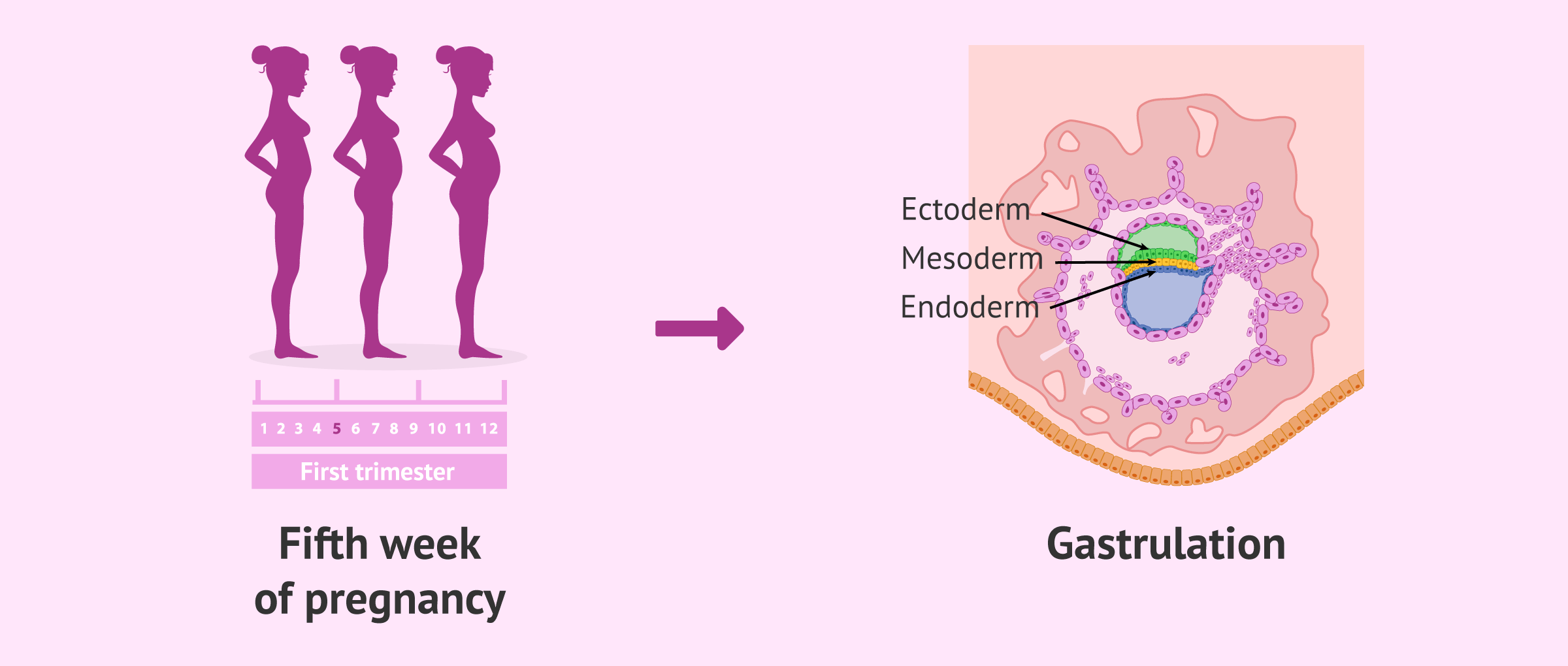
The size of the embryo during the gastrulation process is one or two millimetres, so it cannot even be seen by ultrasound.
On the other hand, it is likely that the woman will begin to feel nausea or other symptoms of pregnancy, but this is due to the increase in the beta-hCG hormone.
Finally, it should be noted that gastrulation is a key process for embryonic development. Any alteration or anomaly during the formation of the three embryonic layers could lead to a halt in the development of the embryo or to the appearance of serious congenital defects.
That is why, it is very important that women start taking care of themselves even before they become pregnant, with a healthy diet and avoiding toxic habits such as tobacco and alcohol.
FAQs from users
What is the neurulation of the embryo?
Neurulation is the embryological process in which the neural tube of the embryo is formed, the precursor structure of the central nervous system. Therefore, the neural tube will give rise to the formation of the brain and the spinal cord.
The neural tube originates from the ectoderm cells that form the neural plate. These cells fold and fuse, forming a cylindrical structure.
The process of neurulation occurs after gastrulation and usually corresponds to the fifth week of pregnancy.
What type of gastrulation occurs in humans?
Vertebrates present different types of gastrulation depending on:
- The shape of the embryo.
- The segmentation mechanism.
- The distribution of yolk.
In the case of human embryos, the process of gastrulation occurs by invagination, also called gastrulation by emboly.
Invagination is the process by which the cells of the inner cell mass (ICM) of the blastocyst fold inward and migrate into the blastocoel, giving rise to the first two embryonic layers: the ectoderm and the endoderm.
In addition to gastrulation by emboly, there are other types such as gastrulation by epiboly, by ingression, by delamination, and by involution. Each of the types of gastrulation mentioned here presents particular characteristics.
Why is gastrulation important?
The importance of gastrulation is due to the fact that it is the beginning from which all the tissues and organs of the human body will be formed. It is, therefore, a key process during embryonic development.
Thanks to the process of gastrulation, the inner cell mass (ICM) of the blastocyst begins to change to give rise to the three embryonic layers: ectoderm, mesoderm, and endoderm.
Suggested for you
Before gastrulation, the embryo undergoes a process of segmentation from being a zygote (egg fertilized with a cell) until it becomes a blastocyst with a multitude of cells. We recommend you read the following post for more information about this: Classification of human embryos according to their quality.
On the other hand, if you want to continue reading about embryonic and fetal development, we recommend you enter the next post: What’s the Difference Between Zygote, Embryo & Fetus?.
Community and Support
At inviTRA we work to make monthly and rigorous information accessible to everyone. If this article has helped you, consider supporting us so we can continue accompanying more people on their journey to parenthood.
References
López-Sánchez C, García-López V, Mijares J, Domínguez JA, Sánchez-Margallo FM, Álvarez-Miguel IS, García-Martínez V. Gastrulación: proceso clave en la formación de un nuevo organismo. Rev Asoc Est Biol Rep 2013; 18(1):29-41. (View)
Solnica-Krezel L, Sepich DS. Gastrulation: making and shaping germ layers. Annu Rev Cell Dev Biol. 2012;28:687-717.
Technau U, Scholz CB. Origin and evolution of endoderm and mesoderm. Int J Dev Biol. 2003;47(7-8):531-9.
Zorn AM, Wells JM. Vertebrate endoderm development and organ formation. Annu Rev Cell Dev Biol. 2009;25:221-51.
FAQs from users: 'What is the neurulation of the embryo?', 'What type of gastrulation occurs in humans?' and 'Why is gastrulation important?'.
Authors and contributors


I find the process of gastrulation quite bizarre… In fact, I’ve never ever heard that word before and I had my son 2 years ago LOL! Very interesting post by the way… Thanks!