Three-dimensional ultrasound or 3D ultrasound was introduced in gynecology in the late 1980s and early 1990s. This insertion represented an evolution in the field of diagnostic imaging, since it offers an amazing resolution and sharpness of the fetus compared to conventional two-dimensional ultrasound.
This type of ultrasound, like the others, is completely safe and does not pose any danger to the health of either the mother or the fetus. Therefore, 3D ultrasound can be performed at any time during pregnancy.
Like any other technique, 3D ultrasound has its benefits and drawbacks. For example, the image offered by 3D ultrasound is sharper and of higher quality than that of 2D ultrasound. However, the time to do a good 3D ultrasound can be quite long.
Provided below is an index with the 9 points we are going to expand on in this article.
- 1.
- 1.1.
- 2.
- 3.
- 4.
- 5.
- 5.1.
- 5.2.
- 5.3.
- 5.4.
- 6.
- 7.
- 8.
- 9.
What is 3D ultrasound?
3D ultrasound is based on the same basic principle as ultrasound, but also incorporates the measurement of fetal volume. This is the main difference with respect to ultrasound performed in two planes or 2D ultrasound.
Thanks to 3D ultrasound it is possible to obtain a static three-dimensional image of the fetus and its structures in sharper color than the black and white of traditional ultrasound. The third plane contained in the 3D ultrasound is the frontal plane, if we are talking about abdominal probes. On the other hand, the third plane would be the transverse plane if vaginal probes are used.
3D ultrasound is usually performed in addition to traditional ultrasound. There is no specific time to perform the 3D ultrasound, although it is recommended to perform this imaging test between the 24th and 32nd week of pregnancy if only a single scan is to be performed.

Today, high-tech equipment is available that offers fast image capture and tracking. Subsequently, a three-dimensional image of the fetus is obtained, which is recorded in the computer software.
With this ultrasound it is possible to differentiate at a few weeks of gestation (12 or 13 weeks of pregnancy), all parts of the fetus' face such as nose, mouth, eyes, ears and others such as the skull, brain and very important the heart. This test helps to detect some malformations or anomalies that may go undetected due to a poor 2D ultrasound image.
How is this ultrasound performed?
The methodology for performing a 3D ultrasound is similar to the procedure for a traditional or 2D gynecological ultrasound. One of the keys to obtaining a good 3D image of the fetus is to first obtain a high-quality 2D image. The 3-D ultrasound is performed in three different phases, but instantaneously, obtaining the image of the fetus in about 3 or 4 seconds:
- First, a fetal volume analysis is performed, both fetal position and 2D fetal size data are collected.
- The second phase consists of the multiplane analysis of all previously collected data.
- In the third phase, the 3D image is reconstructed from the exploration of the 3 planes in space.

In any case, 3D ultrasound can be performed vaginally or abdominally. In addition, it is important that the woman attends the day of the 3D ultrasound with an empty bladder for better observation of the fetus.
Advantages
Since the 3D ultrasound image is much sharper and more informative, it is possible to more accurately detect fetal malformations. Therefore, the main advantage of 3D ultrasound is the ease of diagnosis of fetal alterations, as well as the detection of fetal skin defects such as cleft lip or cleft lip.
Another benefit of 3D ultrasound is that scanning time is reduced. By being able to record images of the fetus, the gynecologist can subsequently perform a more thorough analysis and even consult other experts.

On the other hand, 3D ultrasound makes it possible to measure the volume of the fetus, but it also helps to know the volume occupied by each of the baby's organs. This makes it easier to visualize anomalies in development or organogenesis.
In addition to its usefulness for diagnosis, 3D ultrasound has a great pedagogical value for future parents. The specialist can explain how the baby's development is taking place to the expectant parents after showing the ultrasound images.
Finally, with the 3D ultrasound, the mother and father can have the images of their baby as a souvenir in CD format.
Disadvantages
The main drawback of 3D ultrasound is that a good 2D image is required. Only if you get good 2D images will you get good 3D images.
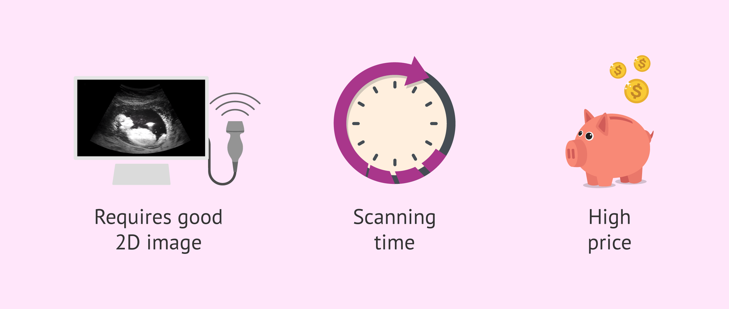
The time taken to get a good image can sometimes be very long. This is because it is a difficult technique to learn and only specialized personnel will be able to perform a good ultrasound. In addition, it is sometimes necessary for the fetus to remain still for the 3D image to be of better quality.
Finally, another possible disadvantage of 3D ultrasound is that its price is higher than that of a normal ultrasound.
Recommendations before performing a 3D ultrasound
3D ultrasound is useful, as long as the conditions at the time of its performance are appropriate. Among the essential requirements when performing a 3D ultrasound are the following:
- Position of the fetus: if the fetus is not facing forward, but is on its back, its face will not be visible.
- Fetal mobility: in 3D ultrasound, fetal movement can interfere with the image sharpness. It is best to wait until the fetus is still before proceeding with the ultrasound.
- Adequate amount of amniotic fluid: too much amniotic fluid can make it difficult to see the baby's face.
- Correct ultrasound transmission: if it is not possible to easily introduce the ultrasound scanner, either due to obesity or abdominal surgeries, the image obtained will not be of high quality.

In addition, if a 4D ultrasound is not going to be done where the baby's movement is of interest, it is important to know that the consumption of food containing glucose could stimulate fetal movement and, therefore, the 3D image would not be as clear.
Finally, it is important to know that 3D ultrasound should not replace any other gestational follow-up ultrasound.
FAQs from users
Is it convenient to perform a 3D ultrasound before IVF?
Yes, 3D ultrasound would provide more information about the uterine cavity. This would allow a better assessment of the anatomy of the uterus, as well as the ovaries and fallopian tubes. In this way and thanks to 3D ultrasound, malformations in the uterus could be detected, such as bicornuate uterus or T-shaped uterus, which could perhaps be overlooked in a traditional ultrasound.
What are the differences between 3D, 4D and 5D ultrasound?
3D ultrasound adds depth to the image, providing a more realistic image. On the other hand, 4D ultrasound also includes the vision of the baby's movement.
The 5D ultrasound is the most advanced and, in addition to contemplating depth and movement, it also improves the resolution through the play of light and shadows. Therefore, 5D ultrasound is the test that offers the best and most realistic images of the baby.
Is it a good time to do a 3D ultrasound in the 19th week of pregnancy?
The general recommendation is to perform the 3D ultrasound between the 22nd and 30th weeks of pregnancy since it will allow a better view of the baby and, therefore, to see its face and not only the bone structure. However, the truth is that you should always consult with a medical specialist when it would be best to do it depending on each situation. In addition, it should be taken into account that the position of the baby is essential for a good visualization.
Is 3D ultrasound safe for the foetus?
Yes, 3D ultrasound is not a dangerous imaging test for the foetus, it is completely safe. Moreover, this test does not pose any danger to the mother's health either.
This is because 3D ultrasound does not emit radiation, but its methodology is based on the application of ultrasound.
Suggested for you
An improved version of 3D ultrasound is 4D ultrasound. If you wish to obtain more information about this imaging test you can continue reading the following link: When is 4D ultrasound performed in pregnancy and what advantages does it offer?
In addition, if you are interested in knowing everything about the controls that are performed during pregnancy, then we invite you to read this article: Prenatal pregnancy control: tests, analyses and ultrasound scans.
Community and Support
At inviTRA we work to make monthly and rigorous information accessible to everyone. If this article has helped you, consider supporting us so we can continue accompanying more people on their journey to parenthood.
References
B Tutschek. 3D prints from ultrasound volumes. Ultrasound Obstet Gynecol. 2018 Dec;52(6):691-698. doi: 10.1002/uog.20108 (View)
Lara Spalldi Barišić, Milan Stanojević, Asim Kurjak, Selma Porović, Ghalia Gaber. Diagnosis of fetal syndromes by three- and four-dimensional ultrasound: is there any improvement. J Perinat Med. 2017 Aug 28;45(6):651-665. doi: 10.1515/jpm-2016-0416 (View)
Linde S Hesse, Moska Aliasi, Felipe Moser; INTERGROWTH-21(st) Consortium; Monique C Haak, Weidi Xie, Mark Jenkinson, Ana I L Namburete. Subcortical segmentation of the fetal brain in 3D ultrasound using deep learning. Neuroimage. 2022 Jul 1;254:119117. doi: 10.1016/j.neuroimage.2022.119117. Epub 2022 Mar 21 (View)
R L Schild, H Plath, C Hofstaetter, M Hansmann. Diagnosis of a fetal mesoblastic nephroma by 3D-ultrasound. Ultrasound Obstet Gynecol. 2000 Jun;15(6):533-6 (View)
S A Chen, C S Ong, N Hibino, A A Baschat, J R Garcia, J L Miller. 3D printing of fetal heart using 3D ultrasound imaging data. Ultrasound Obstet Gynecol. 2018 Dec;52(6):808-809. doi: 10.1002/uog.19166 (View)
S Yagel, S M Cohen, I Shapiro, D V Valsky. 3D and 4D ultrasound in fetal cardiac scanning: a new look at the fetal heart. Ultrasound Obstet Gynecol. 2007 Jan;29(1):81-95. doi: 10.1002/uog.3912 (View)
FAQs from users: 'Is it convenient to perform a 3D ultrasound before IVF?', 'What are the differences between 3D, 4D and 5D ultrasound?', 'Is it a good time to do a 3D ultrasound in the 19th week of pregnancy?' and 'Is 3D ultrasound safe for the foetus?'.
Author

Find the latest news on assisted reproduction in our channels.
