Time-lapse technology, in the context of assisted reproductive technologies, is used to improve the results of In Vitro Fertilization (IVF) cycles. The main advantage of this method is that it allows us to monitor embryo development with videos fully, while they are still in the incubator.
By doing this, it is possible to select the embryos with the highest implantation potential. Some of the most commonly used time-lapse systems today are EmbryoScope, Geri and Eeva.
Provided below is an index with the 9 points we are going to expand on in this article.
- 1.
- 2.
- 2.1.
- 3.
- 4.
- 5.
- 5.1.
- 5.2.
- 5.3.
- 5.4.
- 5.5.
- 6.
- 7.
- 8.
- 9.
What is time-lapse technology?
Time-lapse is an imaging technique used to create videos using a series of pictures taken at particular time intervals. It allows us to see in greater detail the development that occurs in certain intervals, which take place so slowly that are invisible to the naked eye. Thanks to time-lapse technology, we can see them more rapidly and in full detail.
One of the most remarkable advancements achieved in the past few years in the field of Assisted Reproduction is precisely the use of this technology to monitor embryo development in vitro.
Applied to this context, time-lapse technology takes pictures of embryos at different time intervals (every 10 or 20 minutes, for instance), from different viewpoints. With the pictures obtained, this technology creates videos that allows the embryologist to see the entire development process of each embryo individually or collectively, depending on the case.

In short, thanks to time-lapse technology, today we can see what occurs at each stage of embryo development—from the moment ICSI is carried out until they are transferred, frozen, or thrown away.
Advantages over conventional methods
One of the most important steps of IVF cycles is embryo selection. Being capable of choosing the ones with the highest chance of implanting is a key factor to determine the success rate of a cycle.
To date, embryos were evaluated considering morphological parameters only, that is, the only aspects observed were the shape or appearance of each embryo at a particular moment of the day of development.
Some of the morphological parameters typically examined were size and cell symmetry, number of cells, cleavage rate... More on this story: Embryo Classification.
The main limitation of traditional methods was that the evaluation of embryos was conducted only at particular stages of development. In other words, we gather static information from a dynamic process. Subsequently, we miss material information about the embryo development process.
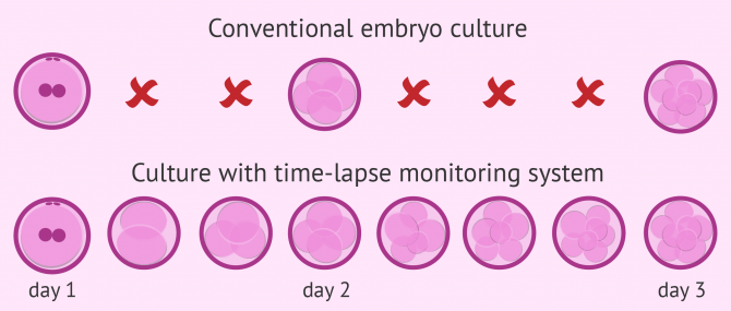
Furthermore, evaluating the quality of embryos under the conventional microscope is not a recommended practice, as quality diminishes each time the embryos leave the incubator.
Thanks to time-lapse technology, we can monitor embryo development without taking the embryos out of the incubator, thereby maintaining them in ideal culture conditions.
What are morphokinetic parameters of embryos?
The development of this technology has led to the emergence of a large amount of new information that was previously inaccessible through traditional microscopic evaluation.
In addition, time-lapse systems also provide the exact timing of specific morphological changes in the embryo. Applying time measurements to morphological evaluation has given rise to a new science: morphokinetics.
Some of the morphokinetic parameters include, for example, the exact time at which the five-cell division occurs or the onset of blastocyst transformation. Other morphokinetic parameters are:
- tPna: time at which the pronuclei appear.
- CC2: duration of the second cell cycle, when embryos progress from 2 to 3 cells.
- CC3: duration of the third cell cycle, transitioning from 3 to 5 cells.
- S2: synchrony of the second cell cycle.
- S3: synchrony of the third cell cycle.

Most time-lapse systems have an integrated algorithm which, based on the collection of these data, can predict which embryos are most likely to implant.
Thanks to this non-invasive method of embryo selection, it is possible to identify which embryos have a higher probability of resulting in pregnancy through the analysis of these implantation-predictive morphokinetic parameters.
Types of time-lapse monitoring systems
Nowadays, the most common time-lapse monitoring systems available on the market can be classified into two categories:
- Incubators with incorporated image systems: EmbryoScope®, Geri®, Miri®, CCM (Cell Culture Monitoring System).
- Independent image systems included in conventional incubators: Primo Vision™, Eeva™.

Each type has a series of pros and particularities, mainly related to the quality of the image system used, the selection algorithms, or the data it provides.
For example, EmbryoScope is able to provide information about oxygen consumption by embryos, which can help to select the embryos more accurately.
Does it increase IVF success rates?
Having as much information as possible about embryo development allows the embryologist to select them with more precision, boosting the implantation rate per transfer.
Also, this improvement favors Single Embryo Transfers (SETs) without affecting the outcomes of IVF, and most importantly, diminishing the risk of multiple pregnancy.
Here is why you should always opt for a single embryo transfer to reduce the risks of IVF: How Many Embryos to Transfer.
A number of studies have focused on determining whether time-lapse monitoring systems actually improve the success rates of reproductive technologies. Indeed, several research studies have proved that this technology increases the pregnancy success rates of IVF.

However, more studies that confirm these results are needed yet. Also, there is an increasing need to determine whether these improvements are due to an optimization of culture conditions, to the use of more accurate embryo selection procedures, or to a combination of both.
There is no doubt, in any case, that time-lapse technology has clear potential to improve IVF success rates, as well as for introducing new expertise in the field of Embryology.
FAQs from users
What are the benefits of Time-Lapse IVF incubators?
Time-lapse incubators have an image capture technology that allows us to generate a film of the entire development of the embryos so that we can analyze the embryonic kinetics.
This allows us to observe the embryo division without removing the embryos from the incubator at any time so that they are not subjected to the temperature changes that occur during this process. This improvement in culture conditions helps more embryos to reach the blastocyst stage.
Time-lapse technology allows us to improve embryo selection, since we will not only rely on specific observation on days 1, 2, 3 and 5 of development, but we will also have information about everything that has happened during the development of the embryo, the times and the way in which these processes have taken place. This makes it easier for us to choose the embryo to be transferred to the uterus.
Is there any difference between the success rates of incubators with a time-lapse system incorporated and independent image systems?
Time-lapse embryo incubators have received much attention since their initial application. However, time-lapse or any imaging system only provides the capability of reviewing the process of embryo development. While it might be able to predict embryo development from the cleavage stage to the blastocyst stage, time-lapse technology has not proven to increase pregnancy success rates over standard IVF incubators.
Can I see the video recording of my embryos?
Yes, once you finish your IVF cycle, your clinic can provide you the DVD with the development of your embryos if you want it. Some labs allow patients to see the development of their embryos live at home.
If you see a poor-quality embryo in a video, do you automatically discard it?
Considering its stage and development stage, the embryologist will determine which one has the highest implantation potential. Even though class C embryos (low quality) have a limited implantation chance, some are able to cause a pregnancy. For this reason, if an embryologist considers that there are chances for it to implant and there are no other embryos of better quality, it will not be discarded but chosen for the transfer.
Will my IVF cycle be more expensive if they use time-lapse technology?
Currently, the main disadvantage of time-lapse technology is that the cost of embryo culture with this system is higher than with conventional methods. For this reason, the cost of IVF may increase if this technology is used. The cost varies from clinic to clinic, though.
Suggested for you
Time-lapse monitoring systems are used for embryo selection during the embryo culture step of the IVF process. Want to learn more about it? Check this out: Embryo Culture Media for Human IVF.
On the other hand, and as we have seen above, there exist several types of time-lapse incubators, but the truth is that EmbryoScope has become very popular nowadays amongst fertility clinics worldwide. Read more: What Is EmbryoScope?
Community and Support
At inviTRA we work to make monthly and rigorous information accessible to everyone. If this article has helped you, consider supporting us so we can continue accompanying more people on their journey to parenthood.
References
Alpha scientists in reproductive medicine and ESHRE special interest group of embryology: The Istanbul consensus workshop on embryo assessments: proceedings of an expert meeting. Hum Reprod 2011, 26:1270–1283.
Borini A, Lagalla C, Cattoli M, Sereni E, Sciajno R, Flamigni C, Coticchio G: Predictive factors for embryo implantation potential. Reprod Biomed Online 2005, 10:653–668.
Chamayou S, Patrizio P, Storaci G, Tomaselli V, Alecci C, Ragolia C, Crescenzo C, Guglielmino A: The use of morphokinetic parameters to select all embryos with full capacity to implant. J Assist Reprod Genet 2013, 30:703–710.
Conaghan J, Cjhen AA, Willman SP, Ivani K, Chenette PE, Boostanfar R, Baker VL, Adamson GD, Abusief ME, Gvakharia M, Loewke KE, Shen S: Improving embryo selection using a computer-automated time-lapse image analysis test plus day 3 morphology: results from a prospective multicenter trial. Fertil Steril 2013, 100:412–419.
Kahraman S, Cetinkaya M, Pirkevi C, Yelke H, Kumtepe Y: Comparison of blastocyst development and cycle outcome in patients with eSET using either conventional or time lapse incubators. A prospective study of good prognosis patients. J Reprod Stem Cll Biotechnol 2012, 3(2):55–61
Kaser DJ, Racowsky C: Clinical outcomes following selection of human preimplantation embryos with time-lapse monitoring: a systematic review. Hum Reprod Update 2014, 20:617–631.
Kirkegaard K, Agerholm IE, Ingerslev HJ: Time-lapse monitoring as a tool for clinical embryo assessment. Hum Reprod 2012, 27:1277–1285.
Kirkegaard K, Hindkjaer JJ, Grondahl ML, Kesmodel US, Ingerslev HJ: A randomized clinical trial comparing embryo culture in a conventional incubator with a time-lapse incubator. J Assist Reprod Genet 2012, 29:565–572.
Kirkegaard K, Kesmodel US, Hindkjaer JJ, Ingerslev HJ: Time-lapse parameters as predictors of blastocyst development and pregnancy outcome in embryos from good prognosis patients: a prospective cohort study. Hum Reprod 2013, 28:2643–2651.
Kovacs: Embryo selection: the role of time-lapse monitoring. Reproductive Biology and Endocrinology 2014 12:124.
Lemmen JG, Agerholm I, Ziebe S: Kinetic markers of human embryo quality using time-lapse recordings of IVF/ICSI-fertilized oocytes.
McLernon DJ, Harrild K, Bergh C, Davies MJ, de Neubourg D, Dumoulin JC, Gerris J, Kremer JA, Martikainen H, Mol BW, Norman RJ, Thurin-Kjellberg A, Tiitinen A, van Montfoort AP, van Peperstraten AM, Van Royen E, Bhattacharya S: Clinical effectiveness of elective single versus double embryo transfer: meta-analysis of individual patient data from randomized trials. BMJ 2010, 341:6945.
Meseguer M, Herrero J, Tejera A, Hilligsoe KM, Ramsing N, Remohi J: The use of morphokinetics as a predictor of embryo implantation. Hum Reprod 2011, 26:2658–2671.
Meseguer M, Rubio I, Cruz M, Basile N, Marcos J, Requena A: Embryo incubation and selection in a time-lapse system improves pregnancy outcome compared with standard incubator: a retrospective cohort study. Fertil Steril 2012, 98:1481–1491.
Mio Y, Maeda K: Time-lapse cinematography of dynamic changes occurring during in vitro development of human embryos. Am J Obstet Gynecol 2008, 199:660.e1–660.e5.
Practice Committee of the Society for Assisted Reproductive Technology and Practice Committee of the American Society for Reproductive Medicine: Elective single-embryo transfer. Fertil Steril 2012, 97:835–842.
Pribenszky C, Mátyás S, Kovács P, Losonczi E, Zádori J, Vajta G: Case report: pregnancy achieved by transfer of a single blastocyst selected by time-lapse monitoring. Reprod Biomed Online 2010, 21:533–536
Rubio I, Galán A, Larreategui Z, Ayerdi F, Bellver J, Herrero J, Meseguer M: Clinical validation of embryo culture and selection by morphokinetic analysis: a randomized, controlled trial of the EmbryoScope. Fertil Steril 2014, 102(5):1287-1294.e5
Rubio I, Kuhlmann R, Agerholm I, Kirk J, Herrero J, Escriba M-J, Bellver J, Meseguer M: Limited implantation success of direct-cleaved human zygotes: a time-lapse study. Fertil Steril 2012, 98:1458–1463.
van Montfoort AP, Dumoulin JC, Kester AD, Evers JL: Early cleavage is a valuable addition to existing embryo selection parameters: a study using single embryo transfers. Hum Reprod 2004, 19:2103–2108.
Wong C, Chen AA, Behr B, Shen S: Time-lapse microscopy and image analysis in basic and clinical embryo development research. Reprod Biomed Online 2013, 26:120–129.
FAQs from users: 'What are the benefits of Time-Lapse IVF incubators?', 'Is there any difference between the success rates of incubators with a time-lapse system incorporated and independent image systems?', 'Can I see the video recording of my embryos?', 'If you see a poor-quality embryo in a video, do you automatically discard it?' and 'Will my IVF cycle be more expensive if they use time-lapse technology?'.


